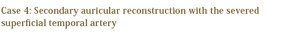
Home > Secondary Auricular Reconstruction #2
Representative Auricular Reconstruction Cases
- Secondary Auricular Reconstruction
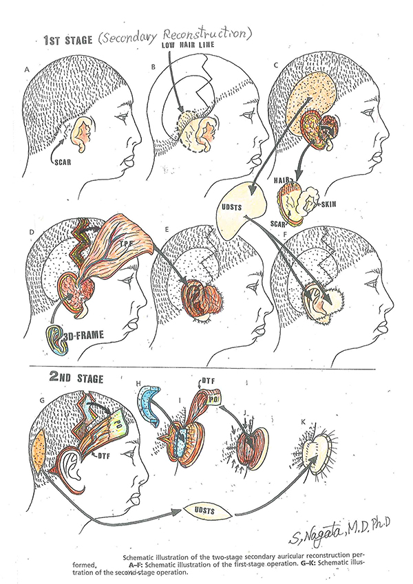
Secondary auricular reconstruction is sought by patients who had auricular reconstruction previously at a different institution that resulted in unfavorable outcomes. Such cases have been rapidly increasing in the last 10 years. It is necessary to remove the cartilage framework from the primary auricular reconstruction, fabricate a new 3-D frame and graft it to its proper anatomical location. This surgery can be more meticulous and complicated at times compared to other reconstruction surgeries due to possible severance of the superficial temporal artery (STA) from the primary auricular reconstruction. However, by using Nagata method for secondary auricular reconstruction with careful surgery planning in advance, favorable and satisfactory results can be attained.
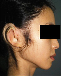
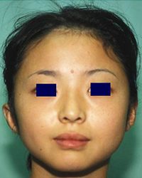
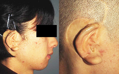
Preoperative appearance
Preoperative appearance
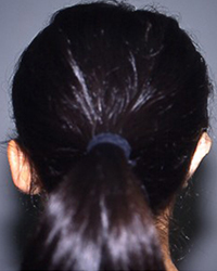
Preoperative appearance
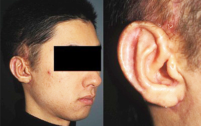
Post-operative appearance with Nagata method
Post-operative appearance with Nagata method
Post-operative appearance with Nagata method
Post-operative appearance with Nagata method
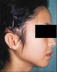
Preoperative appearance:
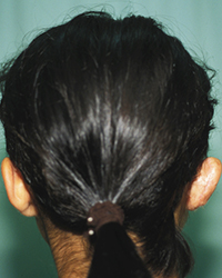
Preoperative appearance reveals the mismatch in color of skin grafted to the conchal vault (a part supposed to be an earhole) and the resorption of the helical rim region of the cartilage framework causing the deformation of the framework in general. In addition, the skin grafted to the back side of auricle was too wide resulting in the abnormal hairline.
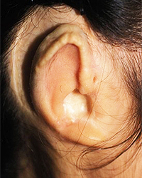
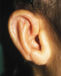
2nd stage post-operative appearance with Nagata method:
The secondary reconstructed auricle with Nagata method is properly projected with the hairline of the mastoid surface (behind the auricle) corrected and well maintained. In the close-up photograph, it appears as if there exists an acoustic meatus beneath the constructed tragus and the color match of the conchal vault with the auricle in general is excellent.
Photos in this blog are shown for your reference for understanding the treatment of microtia. Please understand that surgery results vary depending on each case.
Possible complications following microtia reconstruction surgery
Transient facial palsy, Failure of skin graft due to vascular compromise, Infection resulting in exposure of the 3-D frame, Pneumothorax, Postoperative pneumonia, Suture failure, Alopecia, Decubitus and Others
In the event such complications arise, appropriate action is taken based on the case. Additional or secondary surgery may be required in some situations.
Preoperative appearance Intra-operative appearance
Post-operative appearance with Nagata method
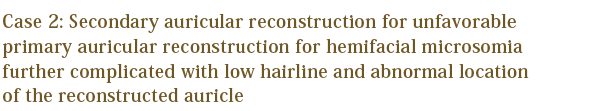
Preoperative appearance:
Preoperative appearance of a hemifacial microsomia case on which the primary auricular reconstruction was performed at a different institution resulted in the patient requesting for a secondary auricular reconstruction with Nagata Method.
Intra-operative appearance:
The intra-operative appearance with the outline reveals the reconstructed auricle located in an improper anatomical location (too anteriorly) due to avoidance of hair-bearing skin and the mismatch in color of skin grafted to the conchal vault (a part supposed to be an earhole). Also, the operative procedure is more complicated due to the low hairline; the auricle to be reconstructed penetrates into the hair-bearing skin.
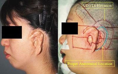
2nd stage post-operative appearance with Nagata method:
All the features of the auricle can be reconstructed. Also, the auricle is in the normal anatomical location and a pseudo acoustic meatus is created. The color match of the skin cover on the conchal vault is excellent.
Photos in this blog are shown for your reference for understanding the treatment of microtia. Please understand that surgery results vary depending on each case.
Possible complications following microtia reconstruction surgery
Transient facial palsy, Failure of skin graft due to vascular compromise, Infection resulting in exposure of the 3-D frame, Pneumothorax, Postoperative pneumonia, Suture failure, Alopecia, Decubitus and Others
In the event such complications arise, appropriate action is taken based on the case. Additional or secondary surgery may be required in some situations.
Preoperative appearance Intra-operative appearance
Post-operative appearance with Nagata method
Photos in this blog are shown for your reference for understanding the treatment of microtia. Please understand that surgery results vary depending on each case.
Possible complications following microtia reconstruction surgery
Transient facial palsy, Failure of skin graft due to vascular compromise, Infection resulting in exposure of the 3-D frame, Pneumothorax, Postoperative pneumonia, Suture failure, Alopecia, Decubitus and Others
In the event such complications arise, appropriate action is taken based on the case. Additional or secondary surgery may be required in some situations.
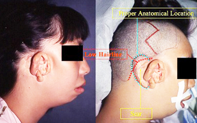
Preoperative appearance:
Preoperative appearance of a hemifacial microsomia case (bilateral) on which the primary auricular reconstruction was performed at a different institution.
Intra-operative appearance:
Intra-operative appearance reveals that auricle reconstructed from the primary reconstruction was located at a great distance from the normal anatomical location (reconstructed on the cheek) as confirmed from the surgical outline and many surgical scars from the primary auricular reconstruction were seen. In addition, this case was further complicated with low hairline.
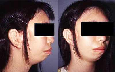
2nd stage post-operative appearance with Nagata method:
All the features of the auricle can be reconstructed. Also, the auricle is in its normal anatomical location with being projected properly.
(Left) Preoperative appearance
(Right) Preoperative appearance, Close-up photo
Post-operative appearance with Nagata method
Preoperative appearance:
Preoperative appearance for an unfavorable primary auricular reconstruction at a different institution with mismatch in color of skin grafted from the groin region. Note that, when the patient reaching puberty, pubic hair growth was seen in the helical rim region and the back side of the auricle where the skin grafted from the groin region.
2nd stage post-operative appearance with Nagata method:
All the features of the auricle can be reconstructed with the auricle being projected properly. The reconstructed auricle has no unwanted hair growth.
Photos in this blog are shown for your reference for understanding the treatment of microtia. Please understand that surgery results vary depending on each case.
Possible complications following microtia reconstruction surgery
Transient facial palsy, Failure of skin graft due to vascular compromise, Infection resulting in exposure of the 3-D frame, Pneumothorax, Postoperative pneumonia, Suture failure, Alopecia, Decubitus and Others
In the event such complications arise, appropriate action is taken based on the case. Additional or secondary surgery may be required in some situations.

For your reference, click here for “Adjustment of ear projection angle for the secondary reconstructed auricle for microtia” conducted on Jan. 16, 2015.